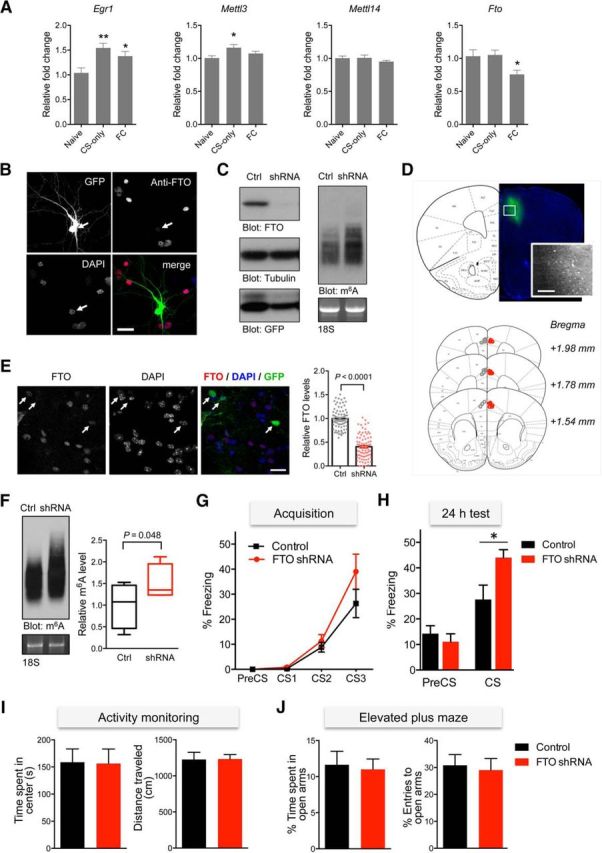
Figure 3.

Targeted knockdown of FTO in the mPFC leads to enhanced fear retention. A, qPCR analyses of Egr1, Mettl3, Mettl14, and Fto in the mPFC following context-exposure (CS-only) or a fear conditioning (FC) paradigm (n = 6–8, one-way ANOVA with Dunnett's test relative to the naive controls, *p < 0.05, **p < 0.01). B, Cortical neurons were transfected with FG12-FTO shRNA, fixed and stained with anti-FTO antibody and DAPI. GFP expression indicates transfected neurons. Scale bar, 25 μm. C, Western blot (left) and m6A immunoblot (right) analyses of cortical neurons transduced with lentivirus particles expressing GFP control (Ctrl) or FTO shRNA. D, Representative image of mouse brain following lentiviral injections into the cingulate (Cgl) and prelimbic (PrL) cortical regions (placement for the control lentivirus indicated in gray and FTO shRNA in red). Scale bar, 200 μm. E, Immunohistochemical analysis of the mPFC region using anti-FTO antibody showing levels of FTO immunoreactivity in shRNA-expressing (GFP-positive) and GFP-negative control cells (n = 80 cells per group from 8 independent slices; Mann–Whitney U test; ****p < 0.0001). Scale bar, 20 μm. F, Representative m6A immunoblot analysis of RNA extracted from the mPFC tissues of mice injected with FG12 control or FTO shRNA lentivirus (n = 4; t test, p < 0.05). G, Cued fear conditioning revealed similar acquisition of fear between groups (control, n = 7; FTO shRNA, n = 8; two-way ANOVA; p = 0.1711). H, The freezing level measured in the same context 24 h after training was significantly higher in the knockdown group (one-way ANOVA with Tukey's post hoc analysis; in CS: control vs FTO shRNA; *p < 0.05). I, An open-field test revealed a comparable duration spent in the center area (left) and total distance traveled (right) by both groups (control, n = 14; FTO shRNA, n = 15). J, An elevated plus maze analysis revealed no differences in the percentage of time spent in (left) or the number of entries (right) to the open arms. Data represent mean ± SEM.
