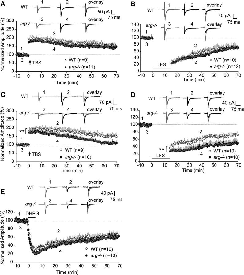
Figure 9.
arg−/− slices exhibit age-dependent alterations in NMDAR-dependent LTP and LTD. A, LTP was recorded in hippocampal slices from WT and arg−/− mice at P21. The magnitude of LTP in arg−/− slices did not differ at any time after TBS at P21. EPSCs were recorded in 20 s intervals at −70 mV for at least 100 min. LTP induction by TBS is indicated by the arrow. Insets show averages of two EPSCs before and after TBS in WT (points 1 and 2) and arg−/− (points 3 and 4) neurons. B, The magnitude of LTD recorded from WT and arg−/− hippocampal slices at P21 was not different. Horizontal bar indicates LTD induction by LFS. Insets show averages of two EPSCs before and after LFS in WT (points 1 and 2) and arg−/− (points 3 and 4) neurons. C, arg−/− neurons at P42 had reduced LTP stability. Insets show averages of two EPSCs before and after TBS in WT (points 1 and 2) and arg−/− (points 3 and 4) neurons. n ≥ 9 slices (four animals). **p < 0.01 vs WT. D, LTD magnitude was significantly enhanced after LFS in arg−/− slices at P42. Insets show the averages of two EPSCs before and after LFS in WT (points 1 and 2) and arg−/− (points 3 and 4) neurons. n ≥ 10 slices (five animals). **p < 0.01 vs WT. E, The magnitude of mGluR-LTD from WT and arg−/− slices at P42 was not different. Horizontal bar indicates DHPG application. Insets show the averages of two EPSCs before and after DHPG in WT (points 1 and 2) and arg−/− (points 3 and 4) neurons. n = 10 slices (five animals).

