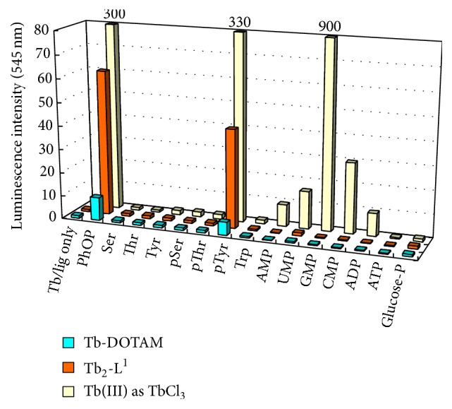
Figure 4.

The luminescence intensity at 545 nm of TbIII-DOTAM (blue bars) and TbIII 2-L1 (red bars) in the presence of various phosphorylated and nonphosphorylated amino acids, nucleoside derivatives, and PhOP (a model compound of pTyr). Conditions: [TbIII complex] = [additive] = 100 μM, pH 7.0 (10 mM HEPES buffer), λ ex = 262.5 nm. For the purpose of comparison, the results using TbIII ion without ligand are also presented (yellow bars). Note that nucleotides (UMP, GMP, CMP, and ADP) showed notable signals and thus selective detection of pTyr was unsuccessful.
