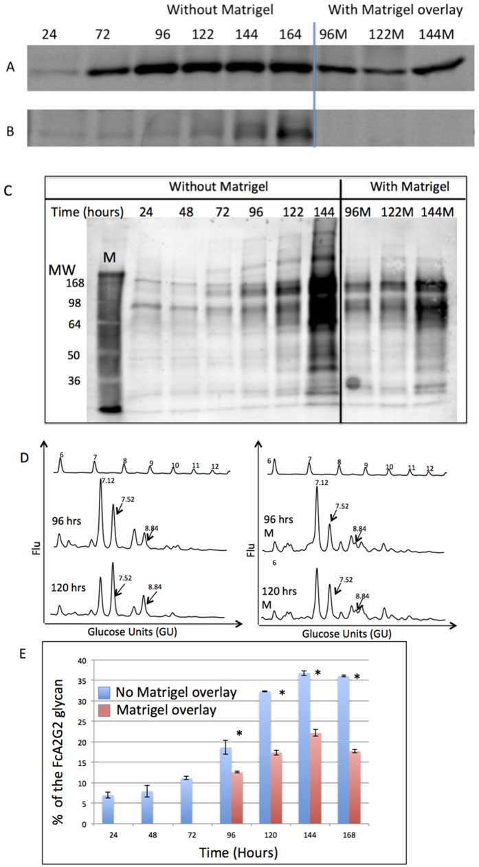
Figure 4. Inhibition of EMT prevents increased levels of fucosylation.
PRH were grown in culture for 60 hours when the process of de-differentiation had begun. At 60 hours, Matrigel was layered on top of the PRH to inhibit the process of de-differentiation. Cells were lysed at the indicated time points and analyzed by immunoblotting to detect (A) Caveolin-1 or (B) Vimentin. (C) Lectin blot using N224Q rAAL to detect fucosylated proteins in lysates from cells prepared as in panels (A,B). (D) N-linked glycan analysis of PRH lysates after 96 and 120 hours of culture without (left) or with (right) Matrigel overlay. The arrows indicate fucosylated glycan peaks that are altered at GU values 7.52 and 8.84. For panel D, glucose values are provided for the major peaks and the GU ladder is provided along the X-axis. Y-axis for these panels represents the fluorescent intensity of glycan. (E) Levels of core fucosylated bi-antennary glycan at the specific times points either without or with Matrigel overlay. In Fig. 4E, the asterisks represent statistical difference (p < 0.05) in samples. For space concerns, Fig. 4A contains cropped images showing the change in Cav or Vim. Analysis of Cav and Vim were performed on the same membrane and thus under the same experimental conditions.

