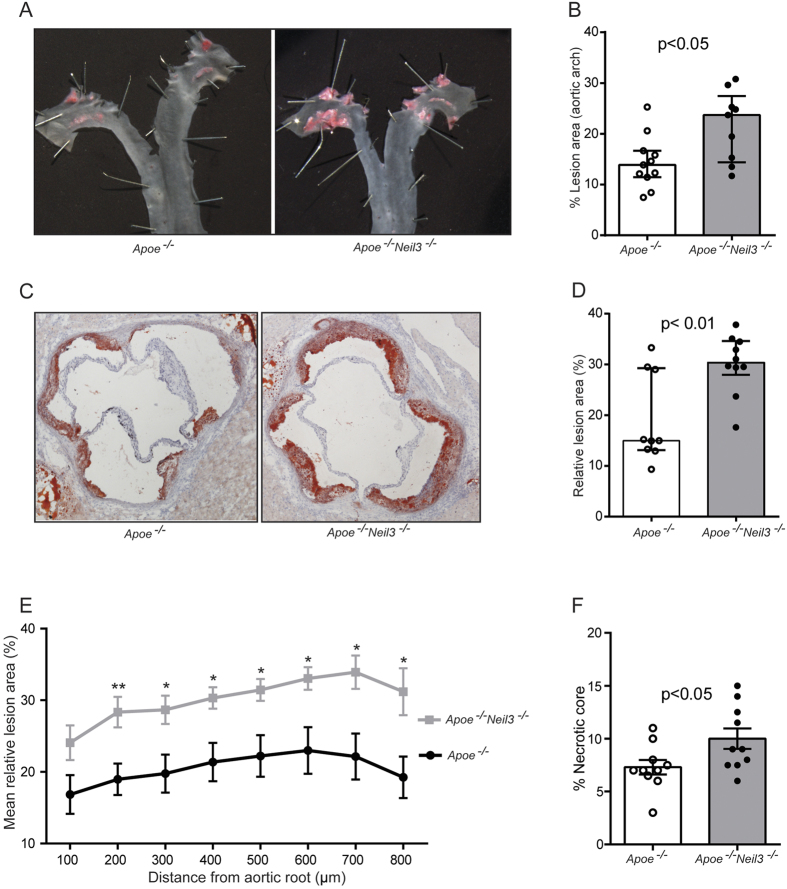
Figure 2. Neil3 deficiency on an Apoe−/− background augments atherosclerosis in male mice fed a high-fat diet.
(A) Representative en face images of the aortic arch stained with Sudan IV. (B) Data show en face % lesion area of Apoe−/− (n = 11) and Apoe−/−Neil3−/− (n = 9) mice. (C) Representative cryosections (10 μm) of the aortic root, stained with Oil Red O and hematoxylin. Original magnification 40X. (D) Relative lesion areas (lesion area/area inside external elastic lamina × 100) in cross-sections of the aortic root, calculated from 8 consecutive sections per mouse at 100 μm intervals in Apoe−/− (n = 9) and Apoe−/−Neil3−/− (n = 10) mice. (E) The graph shows the mean and SEM of relative lesion areas at 8 different positions in the aortic root; n = 9–11 (Apoe−/−) and n = 10 (Apoe−/−Neil3−/−), respectively. *p < 0.05 and **p < 0.01 versus Apoe−/− mice. (F) Necrotic core area as percentage of total plaque area in Apoe−/− (n = 10) and Apoe−/−Neil3−/− (n = 10) mice. Data in (B,D,F) are presented as single values, median and interquartile range and were analyzed using Mann-Whitney U test.

