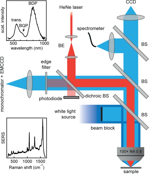
Figure 2.

Schematic Raman/dark-field setup. Pump laser coupled into fully automated microscope with high NA (0.8) 100× objective; Raman emission is isolated with monochromator and EMCCD. Dark-field (DF) scattering spectra are collected by a second fiber-coupled spectrometer. Insets show representative scattering (top) and SERS (bottom) spectra from a single gold nanoparticle on mirror.
