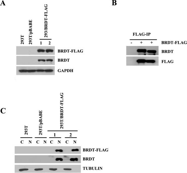
Figure 1. Expression of mouse BRDT in 293T cell lines.
(A) Analysis of ectopically expressed BRDT in selected 293T clones. Approximately 20 μg of whole extract isolated from either 293T, 293T/pBABE, or 293T/BRDT-FLAG cells was analyzed by immunoblotting using either anti-FLAG, BRDT, or GAPDH rabbit polyclonal antibodies. (B) Nuclear extracts from 293T/pBABE and 293T/BRDT-FLAG cells were FLAG-immunoprecipitated, and after extensive washing the retained proteins were analyzed by immunoblotting using anti-BRDT and anti-FLAG antibodies. (C) Immunoblot analysis of BRDT-FLAG nuclear and cytoplasmic expression in one of the stable 293T/BRDT-FLAG cell lines. Detection of tubulin served as a cytoplasmic extract control.

