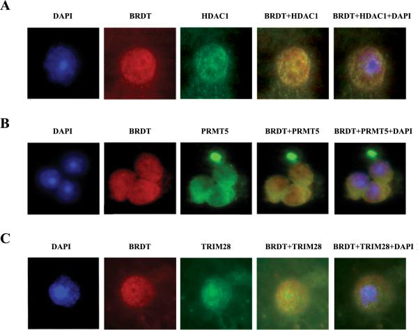
Figure 3. Co-localization of BRDT with HDAC1, PRMT5 and TRIM28 in round spermatids.
(A) Immunofluorescent detection of round spermatids stained for BRDT (red) and HDAC1 (green). Panels (B) and (C) depict the staining for BRDT and PRMT5 and TRIM28, respectively. Nuclei were counterstained with DAPI (blue). Photomicrographs were taken at 100× magnification.

