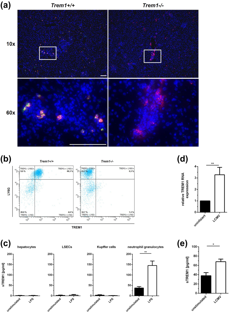
Figure 4. TREM1 expression in LCMV-infected livers of mice by neutrophils.
(A) Frozen liver sections of C57BL/6 mice 9 days after infection with LCMV were stained for TREM1 (green) and Ly6G (red); nuclei were stained with Hoechst 33258 (blue). Co-expression of Ly6G (red) by TREM1-expressing cells (green) in livers of LCMV-infected mice confirmed that TREM1-expressing cells are neutrophils. Scale bar represents 100 μm. (B) Flow cytometric analysis of TREM1 and LY6G expression on neutrophil granulocytes isolated from Trem1+/+ und Trem1−/− C57BL/6 mice 9 days after infection with LCMV. (C) Concentrations of sTREM1 in culture supernatants of primary hepatocytes, liver sinusoidal endothelial cells (LSECs), Kupffer cells or neutrophils 24 hours after isolation from Trem1+/+ C57BL/6 mice and stimulation with LPS (10 ng/ml). Shown are mean values ± SEM. **p < 0.01. (D) Relative TREM1 RNA expression in neutrophils 6 hours after isolation from Trem1+/+ C57BL/6 mice and stimulation with LCMV WE (MOI 5). Shown are mean values ± SEM; **p < 0.01. (E) Release of sTREM1 by neutrophils into cell culture supernatant determined 24 hours after isolation from Trem1+/+ C57BL/6 mice and stimulation with LCMV WE (MOI 5). Shown are mean values ± SEM. **p < 0.01.

