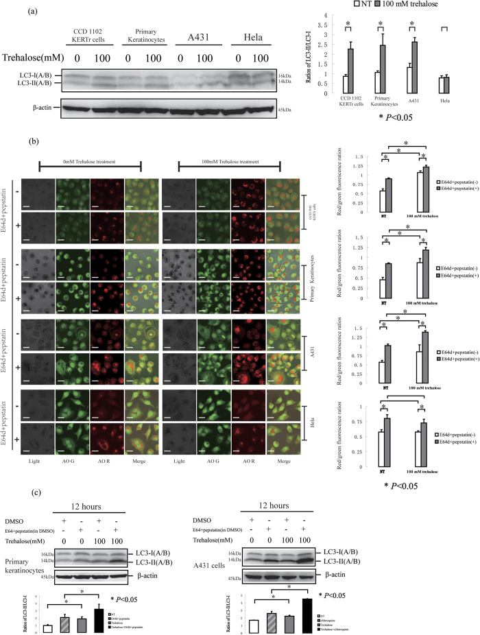
Figure 4. CCD 1102 KERTr cells (keratinocytes transformed HPV 16 E6/E7 oncogenes), human primary keratinocytes, A431 cells and HeLa cells were treated with 100 mM trehalose for 12 hours.
(a) The expression of LC3 protein was determined by western blotting, and then the ratios of LC3-II/LC3-I was calculated for statistical analysis. β-actin served as a loading control. The cells were imaged for AO staining assay using a laser scanning confocal microscope. (b) The means of red/green fluorescence ratios for individual cells in three independent experiments were determined. (c) Human primary keratinocytes or A431 cells were treated with or without 100 mM trehalose in the presence or absence of E64d and pepstatin. The levels of LC3 protein were determined by western blotting, and the ratios of LC3-II/LC3-I was calculated. β-actin served as a loading control. The data were shown as means ± SD of three independent experiments, and representative figures were shown. Bars = 20 μm.

