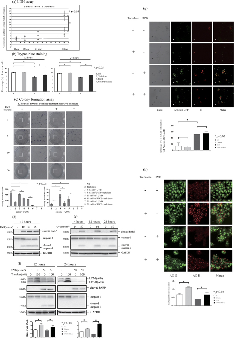
Figure 7. HaCaT cells were treated with or without 50 mJ/cm2 UVB radiation, and then the cells were cultured in the presence or absence of 100 mM trehalose for 4, 12, 24 and 48 hours respectively.
Supernatants of the cells culture medium were performed by LDH assay (A). Cytotoxicity level values (percentage of cell death) were shown as means ± SD of three independent experiments. HaCaT cells were treated with or without 50 mJ/cm2 UVB radiation, and then the cells were cultured in the presence or absence of 100 mM trehalose. Then, the cells were performed by trypan blue staining (B) or by colony formation assay (C). The percentages of survival cells (B) or colony formation counts (C) were shown as means ± SD of three independent experiments. (D–F) Western blotting was used to determine the levels of apoptosis molecular markers (cleaved PARP and caspase-3) and LC3 protein. The cells were imaged for AO staining assay or Annexin V-EGFP apoptosis detection using a laser scanning confocal microscope. The percentages of HaCaT cells marked with Annexin-EGFP and PI were calculated (G). The means of red/green fluorescence ratios for individual cells were determined (H). The means ± SD represented from three independent experiments, and representative figures were shown. Bars = 20 μm.

