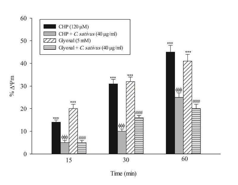
Fig. 5 .

Effect of C. sativus on mitochondrial membrane potential decline during CHP and glyoxal induced hepatocyte injury. Mitochondrial membrane potential was determined as the difference in mitochondrial uptake of the rhodamine 123 between control and treated cells. Our data were shown as the percentage of mitochondrial membrane potential collapse (%ΔΨm) in all treated (test) hepatocyte groups. Values are expressed as the mean±SD of three separate experiments (n=3). *** p < 0.001compared with control hepatocytes in the same time; ФФФ p < 0.001 compared with CHP treated hepatocytes and ### p < 0.001 compared with glyoxal treated hepatocytes.
