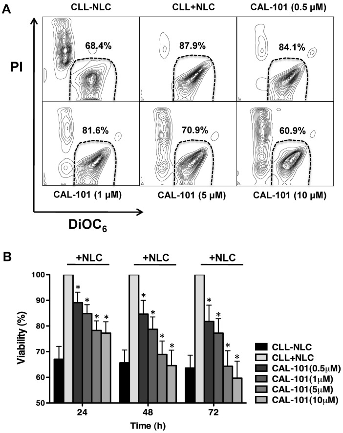
Figure 3.
CAL-101 antagonizes NLC-mediated CLL cell survival. (A) CLL cells were cultured alone (control), cocultured with NLCs in medium alone or in medium containing various concentrations of CAL-101. Displayed are representative contour plots that depict CLL cell viability after 48 hours and after staining with DiOC6 and PI (horizontal and vertical axes, respectively). The viable cell population is characterized by bright DiOC6 staining and PI exclusion, and is gated in the bottom right corner of each contour plot. The percentage of viable cells is displayed above each of these gates. (B) The bar diagram represents the mean relative viabilities of CLL cells cocultured with NLCs compared with CLL cells alone (control) and cocultured with NLCs plus various concentrations of CAL-101. Viabilities of CAL-101–treated samples were normalized to the viabilities of control samples at the respective timepoints (100%). Displayed are the means (± SEM) from 12 different patient samples, assessed after 24, 48, and 72 hours. CLL cell survival in the presence of NCLs was significantly inhibited by CAL-101, with P < .05, as indicated by the asterisks describing the comparison of results from each culture containing CAL-101 to the results from the control culture.

