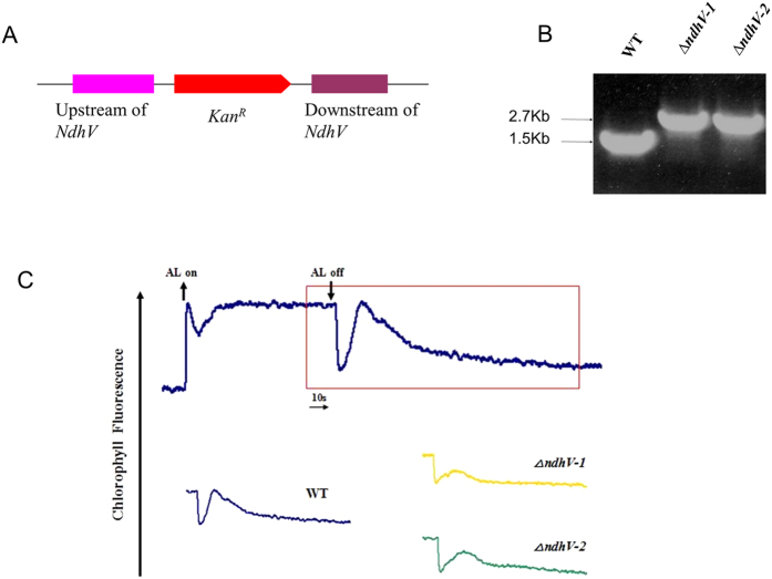
Figure 1. NdhV gene deletion and its effect on NDH-1 activity.
(A) Construction of plasmid to generate NdhV deleted mutant (ΔndhV). Schematic representation of the ΔndhV mutant locus. A kanamycin resistance cassette about 1.2Kb was inserted into the NdhV gene. (B) PCR segregation analysis of the ΔndhV mutant using the ndhV-up-F and ndhV-Dn-R primers (Table S1). (C) Monitoring of NDH-1 activity using chlorophyll fluorescence analysis. The top curve shows a typical trace of chlorophyll fluorescence in the WT of Synechocystis 6803. The cells (OD730 around 0.4) supplemented with 10 mM NaHCO3 were used for the measurement. After the sample was exposed to the actinic light (AL, 100 μmol photons m−2 s−1) for 90 s, AL was turned off, and the transient increase in chlorophyll fluorescence level was recorded, which was used to ascribe NDH-1 activity. The inset shows magnified traces from the box area.

