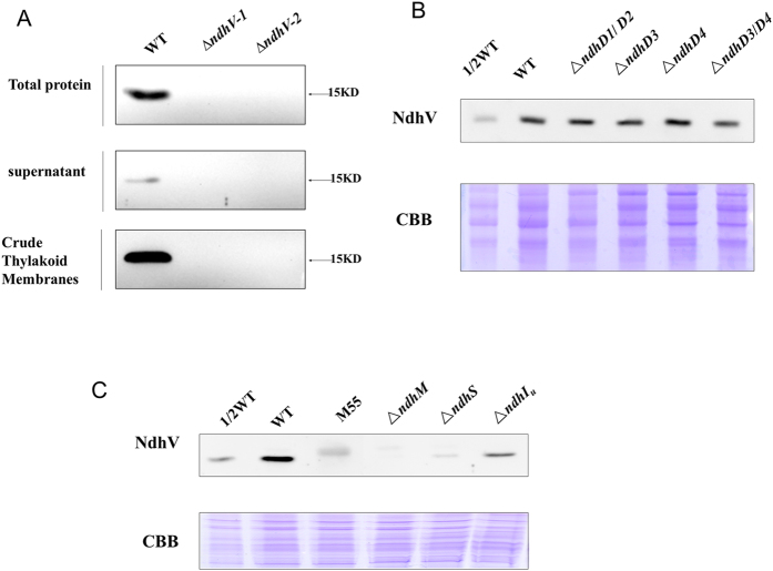
Figure 4. The location of NdhV in WT strain and the effects of mutation of Ndh subunits on NdhV.
(A) Immunodetection of NdhV in the total proteins, supernatant and thylakoid membranes of WT and ΔndhV strains. Total Proteins: The material obtained after broken by glass beads; supernatant, Crude Thylakoid Membranes: supernatant and precipitation after centrifugation of total proteins at 20, 000 × g for 30 min at 4 °C, respectively. (B) Immunodetection of NdhV in thylakoid membranes from WT (including indicated serial dilutions), ΔndhD1/D2, ΔndhD3, ΔndhD4 and ΔndhD3/D4 mutants. Immunoblotting was performed using antibodies against NdhV. Each Lane was loaded with 25 μg total proteins. In the lower panel, a piece of replicated gel stained with Coomassie Brilliant Blue (CBB) was used as a loading control. (C) Immunodetection of NdhV in thylakoid membranes from WT (including indicated serial dilutions), M55, ΔndhM, ΔndhS, ΔndhIU(partly deletion of NdhI) mutants. Immunoblotting was performed using antibodies against NdhV. Each Lane was loaded with 25 μg total proteins. In the lower panel, a piece of replicated gel stained with Coomassie Brilliant Blue (CBB) was used as a loading control.

