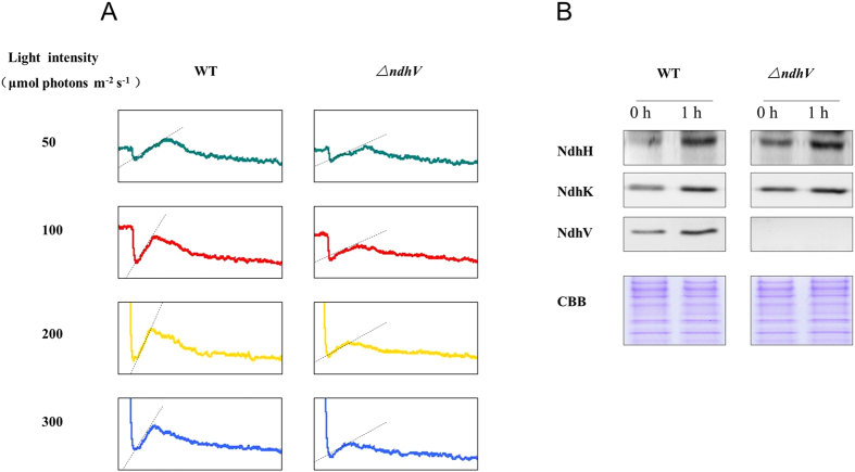
Figure 6. Monitoring of NDH-1 activity of WT and ΔndhV strains under different light intensities.
(A) Monitoring of NDH-1 activity of WT and ΔndhV strains in different light intensities using chlorophyll fluorescence analysis. OD730 of the cells was about 0.3. The cells were exposed to the different actinic light (shown in the figure) for 90 s. Then the actinic light was turned off, and the transient increase in chlorophyll fluorescence level was ascribed to NDH activity. (B) Immunodetection of NdhH, K, V in total proteins of WT and ∆ndhV strains before and after treatment with high light. The cell was cultured to mid-logarithmic phase under normal light (0 h), then the cell cultures were transferred to high light (~200 μmol photons m−2 s−1) for 1 hour (1 h). Immunoblotting was performed using antibodies against NdhH, NdhK and NdhV. Each lane was loaded with 25 μg proteins. In the lower panel, a piece of replicated gel stained with Coomassie Brilliant Blue (CBB) was used as a loading control.

