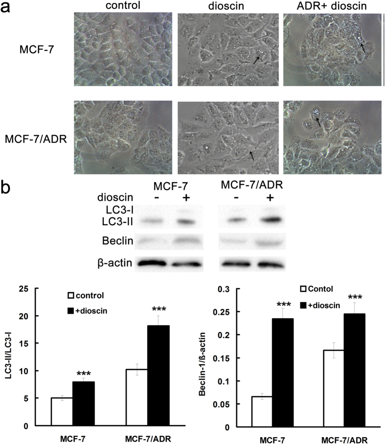
Figure 4. Dioscin induced autophagy in MCF-7 and MCF-7/ADR cells.
(a) Cells were treated with 0.4 μM dioscin for 24 h and cellular morphology was observed by phase-contrast microscopy. Vacuoles in the cytoplasm were marked by arrowhead. Magnification: 400×. LC3-II and beclin-1 protein expression was determined by Western blotting (b). Means ± SD of three experiments are presented.***p < 0.001 versus that obtained in control group.

