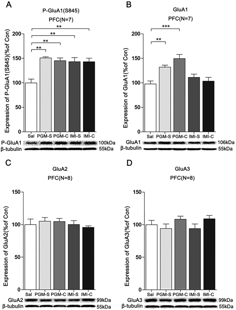
Figure 8. Proteo-β-glucan from Maitake (PGM) evoked a prolonged increase of p-GluA1(S845) (P-GluA1) and GluA1 levels in the prefrontal cortex (PFC).
CD-1 mice were i.p. injected with a high dose of PGM (12.5 mg/kg/day) or imipramine (15 mg/kg/day, IMI) or saline (Sal) for 5 days. Then, the treatment with PGM or imipramine was stopped for 3 days. As shown in the figure, the PGM-S and IMI-S indicated the groups that stopped treatment with PGM or imipramine; PGM-C and IMI-C indicated the groups that continued the treatment. Western blot analyses of the proteins from the PFC were performed with anti-p-GluA1(S845), anti-GluA1, anti-GluA2 or anti-GluA3 antibodies. The number (N) of mice per group is indicated in each individual graph. Data were analysed by one-way ANOVA and presented as the mean ± SE (post hoc Tukey’s test, *p < 0.05, **p < 0.01, ***p < 0.001). The expression levels of (A) p-GluA1(S845), (B) GluA1, (C) GluA2, or (D) GluA3 in the PFC were determined.

