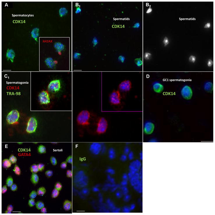
Fig. 2.
Immunofluorescent localization of CDK14 in testicular cells. Color-coding is indicated for each image. Scale bar is 10μm. Nuclei are stained by DAPI (blue). Different cell types have been separated from adult and pubertal testes as described in Section 2. (A) Spermatocytes; insert: double staining for CDK14 and γH2AX (a spermatocyte marker). (B1) Spermatids; (B2) DAPI alone panel of Fig. B1 to visualize different types of spermatid nuclei. (C1) TRA- and CDK14-positive spermatogonia; (C2) CDK14 staining alone of Fig. C1. (D) GC-1 spermatogonia. (E) Mouse immature Sertoli cells. (F) Negative control (rabbit IgG was used instead of primary antibody).

