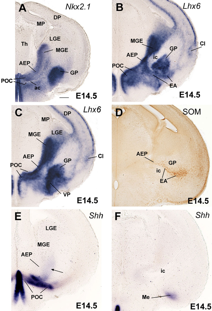
Fig. 2.
Frontal sections through the embryonic telencephalon of different mouse embryos (E14.5) hybridized for Nkx2.1, Lhx6, or Shh, or immunostained for the neuropeptide somatostatin (SOM). Sections shown in A,C,E are at similar frontal levels (at about the level of the anterior commissure; ac), to facilitate comparison of expression patterns. Section shown in B is just caudal to the anterior commissure. Sections shown in D,F are at a more caudal level, when the internal capsule (ic) enters/exists the telencephalic vesicle. Lhx6 expression shows three distinct radial subdivisions in the subpallium: MGE, AEP, and POC. Only the POC shows strong expression of Shh in the ventricular zone (E), which extends laterally, invading the dorsolateral preoptic area and the medial amygdala (Me; F). A band of weak Shh expression extends from the POC domain into AEP/MGE, and may represent tangentially migrating cells (arrow in E). MGE is radially correlated with the globus pallidus (GP). The AEP is sandwiched between MGE and POC. It consists of a thick radial band of Lhx6 expression extending from the subventricular zone to the surface, below the GP. It appears to give rise to the sublenticular part of the extended amygdala (EA). It is also related to a corridor of SOM+ cells (D), which appear to originate in AEP. At this age, Lhx6 is also expressed in cells that migrate tangentially to the cortical/pallial regions (including the cerebral cortex and claustrum). See text for more details. For abbreviations, see list. Scale bar = 200 µm. [Color figure can be viewed in the online issue, which is available at www.interscience.wiley.com.]

