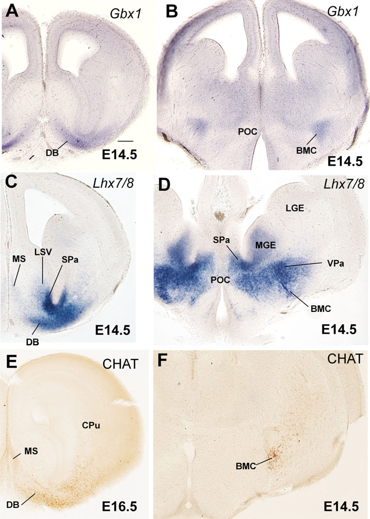
Fig. 4.
Frontal sections through the embryonic telencephalon of different mouse embryos (E14.5, E16.5) hybridized for Gbx1 or Lhx7/8, or immunostained for the enzyme choline acetyltransferase (CHAT, a marker of cholinergic neurons). Sections in A,C,E are at a similar level, where the primordium of the diagonal band nucleus (DB) is observed. Sections in B,D,E are at a similar level behind the anterior commissure, where the primordium of the basal magnocellular nucleus (BMC) is present. Note the correlation between Gbx1 expression and the cholinergic cells of the developing BMC and DB. See text for more details. For abbreviations, see list. Scale bar = 200 µm. [Color figure can be viewed in the online issue, which is available at www.interscience.wiley.com.]

