
Figure 2.
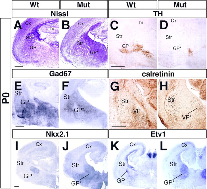
Impairment of GP formation in Arx mutant brains. A, B, P0 Coronal sections stained for Nissl unravel drastic alterations in the cytoarchitecture of the ventral telencephalon and a loss of the GP in the Arx mutants. C, D, TH+ dopaminergic afferent fibers to the GP are lacking in Arx mutants. E, F, GAD67 staining failed to identify the basal nuclei in Arx mutants found in control tissue. G, H, Calretinin typically labels GP interneurons and allows the visualization of the entire GP. In mutant tissue, such a structure is lacking (H). I–L, Molecular markers of GP neurons, such as Nkx2.1 (I, J) and Etv1 (K, L), are not detected in the putative GP location in mutant tissue but appear to be ectopically expressed in a medial–ventral location. Cx, Cerebral cortex; hi, hippocampus; GP*, mutant globus pallidus; Mut, mutant; Str, striatum; VP, ventral pallidum; VP*, mutant ventral pallidum. Scale bars: A (for A, B), C (for C, D), I (for I–L), 200 μm; E (for E, F), G (for G, H), 150 μm.