
Figure 3.
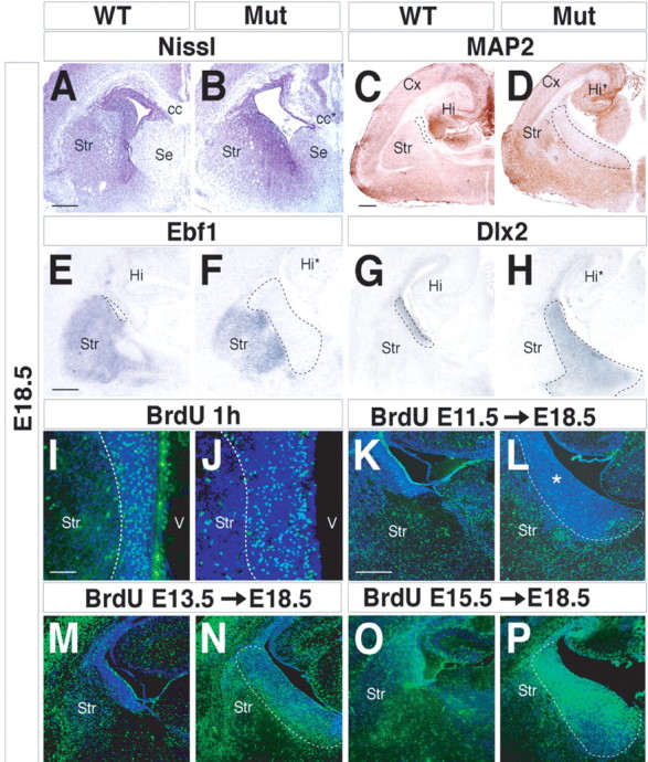
Characterization of the abnormal periventricular area found in Arx mutants. A, B, E18.5 coronal sections stained for Nissl outline the cytoarchitectural defects of the Arx mutant striatum. Marker genes for mature striatal neurons, such as MAP2 (C, D) and Ebf1 (E, F), are not detected in the mutant periventricular area (dashed lines). G, H, Dlx2 transcripts identify the enlarged mutant periventricular region. I, J, Labeling of proliferating cells located within the periventricular area. An expanded area of BrdU-positive cells was observed in mutants (I, J, dashed lines), although without any modification in the number of proliferating cells. K–P, Birth-dating analysis of basal ganglia neurons during development. Pregnant females received a single BrdU injection at E11.5 (K, L), E13.5 (M, N), and E15.5 (O, P), and the location of labeled cells was examined at E18.5. K, L, A single BrdU pulse at E11.5 marks cells distributed throughout the mature compartment both in controls and Arx mutants. *, Expanded mutant SVZ. M–P, Mutant cells labeled at E13.5 and E15.5 are progressively accumulated within the periventricular area (dashed lines) and fail to migrate into the striatal mantle zone. cc, Corpus callosum; cc*, mutant corpus callosum; Cx, cerebral cortex; Hi, hippocampus; Hi*, mutant hippocampus; Mut, mutant; Se, septum; Str, striatum; V, ventricle. Scale bars: A (for A, B), E (for E–H), K (for K–P), 150 μm; C (for C, D), 200 μm; I (for I, J), 100 μm.