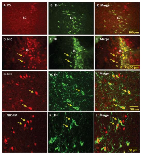
Figure 1.

Fluorescent and laser scanning confocal microscopy images of representative brainstem sections demonstrating nicotine (NIC) and nicotine pyrrolidine methiodide (NIC-PM) activation of noradrenergic neurons of locus coeruleus (LC). Panels A-C: Control data demonstrating the effects of acute intraperitoneal injection of physiological saline (PS) on c-Fos activation of tyrosine hydroxylase (TH)-immunoreactive (IR) cells of LC. Panels D-I: Low power fluorescent (D-F) and high power confocal (G-I) images showing NIC-induced c-Fos IR cells (D, G), TH IR cells (E, H) and merge images of c-Fos with TH IR cells in LC. Panels J-L: High power confocal images showing NIC-PM induced c-Fos IR cells (J), TH-IR cells (K) and merge images of c-Fos with TH-IR cells (L) in LC. Arrows point to representative double-labeled neurons.
