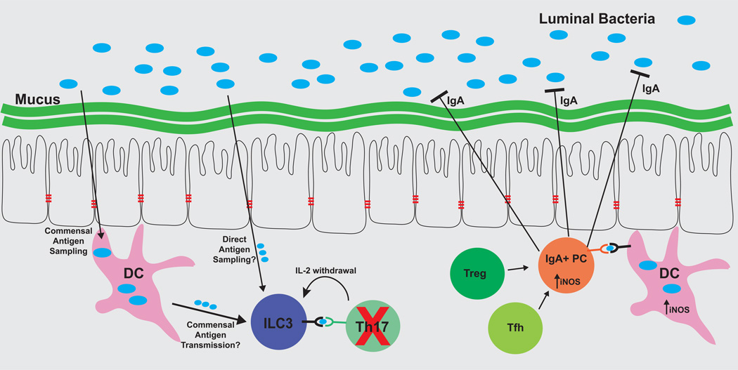
Figure 2. Antigen-dependent host pathways serve both to limit and also to protect commensal bacteria populations.
ILC3 cells and IgA+ plasma cells each respond to commensal bacteria antigens, but ILC3 serves to limit host responses to these antigens, while IgA functions to limit colonization of these bacteria. ILC3 cells acquire antigen through an unknown mechanism, and present commensal bacteria-derived antigens through MHCII, but they do not express co-stimulatory molecules, and instead bind important pro-survival cytokines including IL-2. As a result, ILC3 cells induce apoptosis of T cells reactive to commensal bacteria. In parallel, antigen-specific IgA+ is induced in lymphoid tissues in a DC- and iNOS-dependent manner. T cells can also contribute to antigen-specific IgA responses. Antigen-specific IgA is secreted into the intestinal lumen, where it coats antigenic bacteria and inhibits bacterial colonization of the intestinal epithelium and mucus layers.

