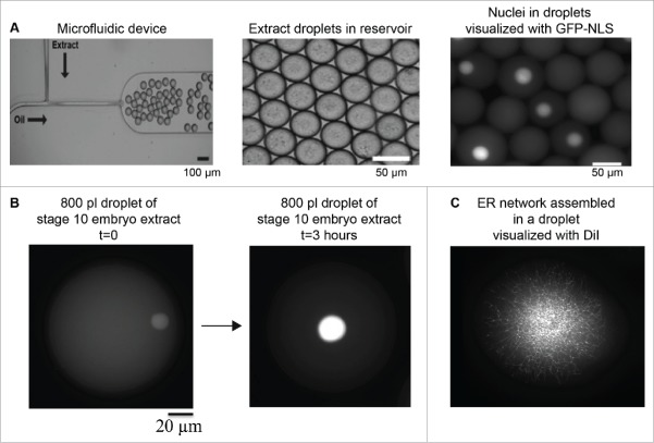
Figure 3.
Microfluidic encapsulation technology to study organelle size scaling. (A) A standard microfluidic T-junction device is shown. At the junction where oil/surfactant and X. laevis egg extract mix, droplets are generated. Droplet size can be tuned by altering device geometry and flow rates. Image courtesy of John Oakey and Jay Gatlin. (B) Large 800 pl droplets containing stage 10 X. laevis embryonic cytoplasm and endogenous nuclei are shown. Nuclei are visualized by import of GFP-NLS. Over the course of ∼3 hours at room temperature, nuclear size expands (our unpublished data). (C) Partially fractionated X. laevis egg extract was encapsulated in a droplet, and ER network formation was visualized with DiI (our unpublished data).

