
Figure 3.
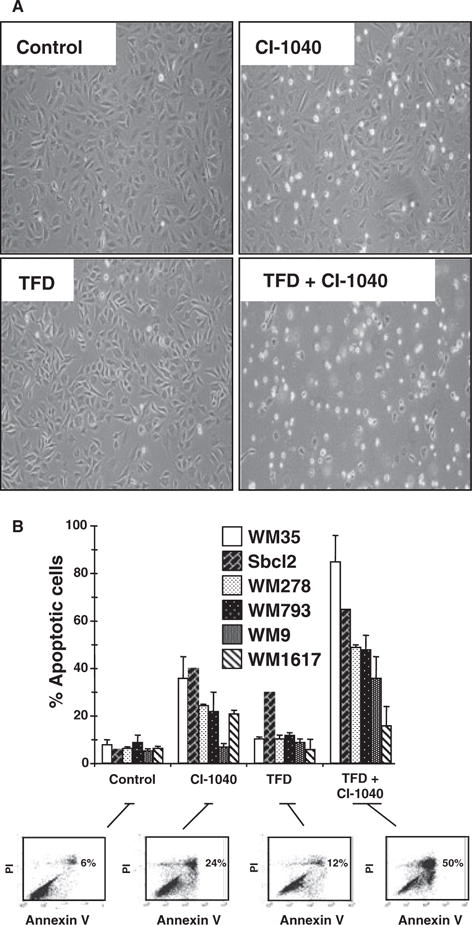
Regulation of apoptosis in human melanoma cells. (A) WM793 cells were treated with 2 μM CI-1040 in the absence or presence of serum for 48 h as indicated. Cells were photographed under phase contrast using a Nikon TMS microscope (magnification 100×). (B) A panel of human melanoma cell lines was subjected to 48 h of TFD in the absence or presence of 2 μM CI-1040 as indicated. Cells were harvested and their viability assessed by Annexin V/propidium iodide co-staining followed by analysis by flow cytometry. Results shown for each cell line are the mean ± SD of at least three individual experiments. Flow cytometry data are shown for one representative experiment for the WM278 cell line.