Figure 5.

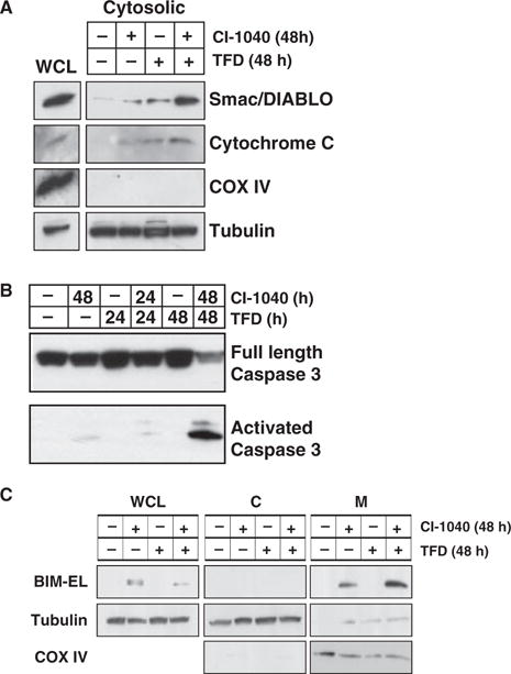
Effect of MEK inhibition and trophic factor deprivation on markers of apoptosis. (A) WM793 cells were treated with 2 μM CI-1040 in the absence or presence of serum for 48 h as indicated at which time cells were harvested. Whole cell and cytosolic lysates (cytosolic) were analyzed by Western blot analysis for the presence of Smac/DIABLO, cytochrome c, the inner mitochondrial marker COX IV and the cytosolic marker β-tubulin. (B) WM793 cells were treated with 2 μM CI-1040 in the absence or presence of serum for 24 or 48 h as indicated. The presence of full length pro-caspase 3 and activated caspase 3 was assessed by Western blot analysis of cell lysates. (C) WM793 cells were treated with 2 μM CI-1040 in the absence or presence of serum for 48 h at which time extracts were prepared. WCL, cytosolic (C) and mitochondrial fractions (M) were prepared as described in Materials and Methods. The presence of BIM-EL, tubulin (cytosolic marker) and COX IV (mitochondrial marker) in each cell fraction was assessed by Western blot analysis of cell extracts.
