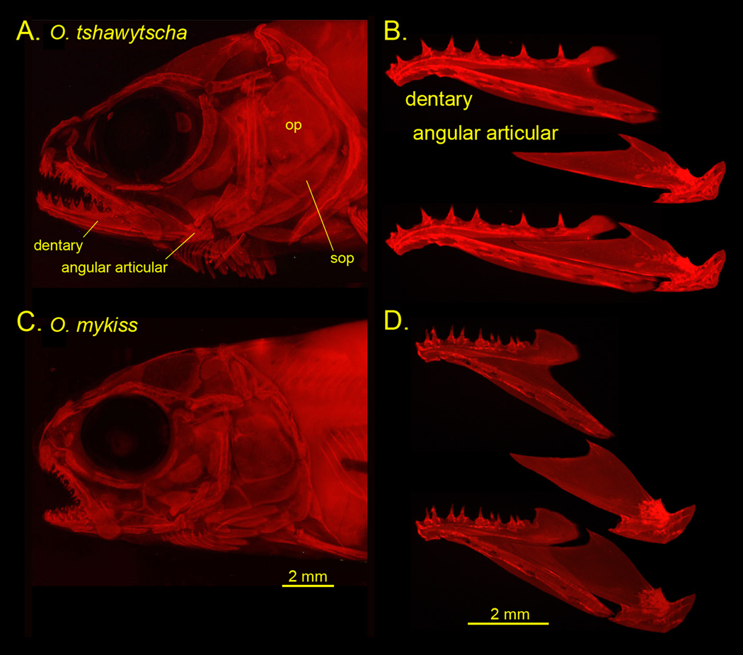
Fig. 2.
A, C, Parr skull preparations, and B, D dissected mandibular bones along with reconstructions of how they fit together. Alizarin Red staining and photographed by epifluorescence. op: opercle, sop: subopercle. The preparations are shown as left side views, with anterior to the left and dorsal up. We follow this convention in the diagrams shown in subsequent figures.

