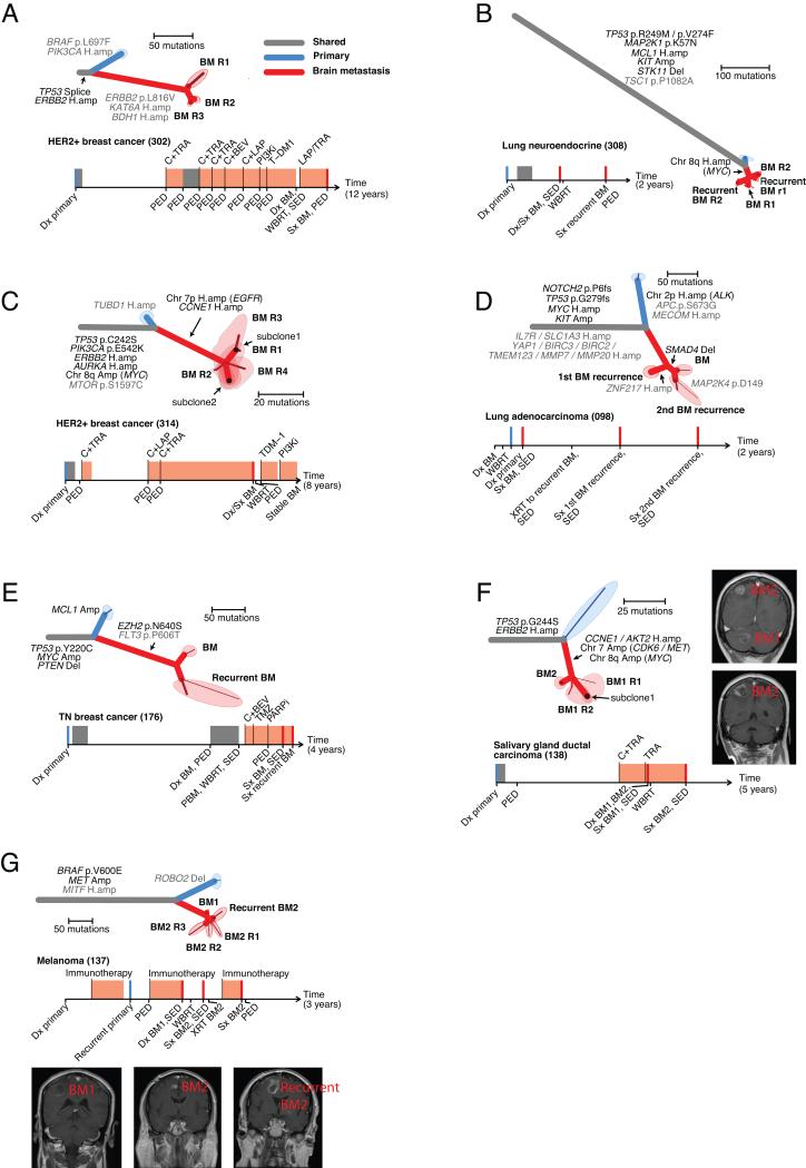
Figure 3. Anatomically and regionally distinct brain metastasis samples share actionable drivers.
A-G. Seven cases for which multiple regionally separated or anatomically distinct brain metastases were sequenced. The samples labeled R1, R2, etc. refer to different regions of the same pathology block. Phylogenetic trees and clinical histories are shown for each case as in Figure 2. C,F., minor subclones shared by > 1 tissue sample were detected (as described in the methods). For these cases, the shared areas denote the tissue samples, and indicate which subclones are present in each sample. F,G. Gadolinium-enhanced MRIs of the sampled brain metastases are shown.

