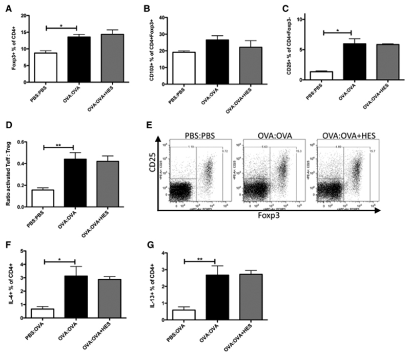
Figure 10. HES at challenge does not affect T-cell accumulation or cytokine production in the lung.
(A-E) Lung tissue cells were prepared from the experiment shown in Figure 8A and stained for CD4, CD25, CD103 and Foxp3 for flow cytometry. Shown are (A) the Foxp3+ proportion of CD4+ cells, (B) the CD103+ proportion of CD4+Foxp3+ cells, (C) the percentage of CD25+ cells among CD4+Foxp3− cells, (D) the ratio of activated effector T cells (CD4+Foxp3−CD25+) to Treg cells (CD4+Foxp3+) and (E) representative flow cytometry plots of CD4+ cells showing Foxp3 verses CD25. Results are representative of 2 repeat experiments with 4-6 mice per group. (F, G) In the second experiment, lung tissue cells from 5 individual mice were stimulated with PMA, Ionomycin and Brefeldin A for 4 hours, and stained for CD4 and intracellular (F) IL-4 and (G) IL-13 by flow cytometry. Error bars are SEM. *p<0.05, **p<0.01, one-way ANOVA, with a Tukey’s post test.

