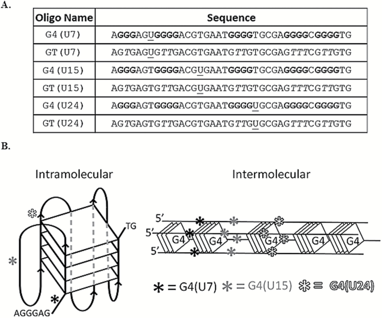
Figure 1.
Sequences used and diagrams of G4 structures. (A) The name (left) and sequence (right) for each oligonucleotide are shown. G4 and GT indicate oligonucleotides capable or incapable of adopting G4, respectively. The nucleotide position of the uracil, U7, U15 and U24 relative to the 5′ end is indicated within each oligonucleotide name. The positions of uracil bases within each sequence are underlined and guanine repeats are bolded. Substitution of thymine for guanine interrupts G4 folding potential, shown with an italicised ‘T.’ (B) Diagram depicting intramolecular, left, or intermolecular, right, G4 conformations and the relative positions of each uracil (asterisks). All three uracil positions are depicted on a single G4 conformation for convenience. Diagrams are not scaled models. Actual G4 conformations in solution are predicted to be a mixture of intra and inter-molecular species, but U7 and U24 will be adjacent to a G-tetrad.

