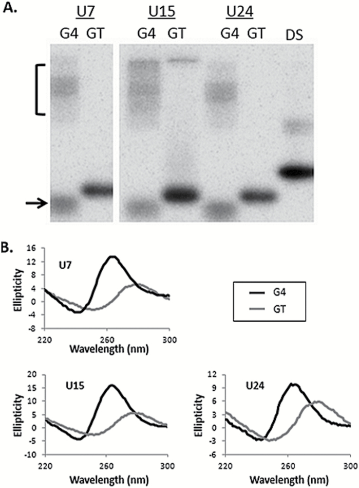
Figure 2.
Detection of G4 formation. (A) Phosphorimages showing native PAGE resolution of 5′ 32P end labelled G4 and GT oligonucleotides in neutral pH and 100mM KCl. The name of each oligonucleotide is shown at the top. Oligonucleotides where guanine repeats are interrupted by thymine to prohibit G4 formation (GT) migrate predominately as a single species. Guanine repeat oligonucleotides (G4) migrate faster (arrow) and slower (bracket) than each control (GT). All oligonucleotides are equal nucleotide length and migration of G4(U24) duplex DNA (DS) is shown as a reference for double-stranded DNA. (B) CD of each G4 and GT uracil-containing oligonucleotide. Ellipticity is shown on the Y axis, wavelength (nm) on the X axis. Spectra for uracil-containing oligonucleotides with guanine repeats (G4) are in black, repeat interrupted (GT) oligonucleotides in grey. All oligonucleotides were assayed in 100mM KCl solution at pH 7.6.

