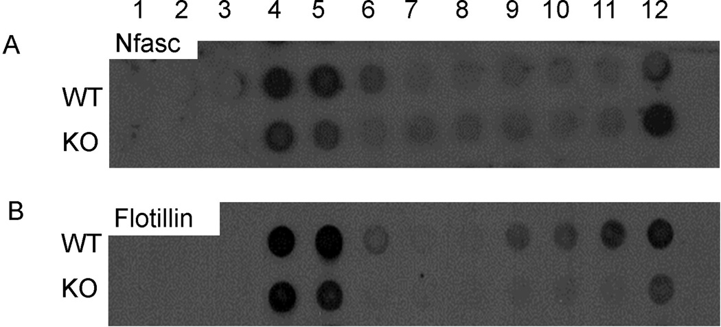
Figure 5. Myelin neurofascin from CST KO spinal cords is isolated from the subfraction with the highest sucrose concentration.
Purified myelin was isolated from spinal cords of CST WT and KO mice and subfractionated by a sucrose gradient. Subfractions were probed with antibodies against in neurofascin (A) and flotillin (B), a putative indicator of membrane raft fractions. Consistent with previous reports, neurofascin from the WT accumulated in the higher fractions (lower sucrose concentrations) of the gradient. In contrast, a dramatic shift was observed with regard to the distribution of neurofascin from the CST KO sample as neurofascin accumulated in the lowest fraction (fraction 12). (B) In the CST WT samples, flotillin was observed in both the higher fractions (fractions 4 and 5) and the lower fractions (fractions 9–12); however, in the CST KO samples, flotillin was primarily observed in fractions 4 and 5 with a small portion of the protein accumulating in fraction 12.

