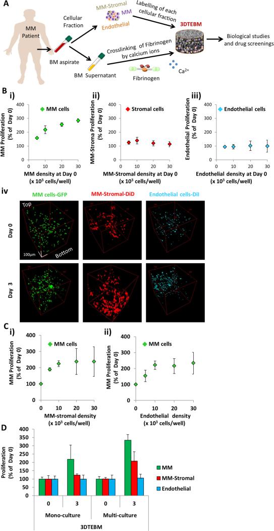
Figure 1. 3DTEBM cultures allow MM cell proliferation and interaction with accessory cells.
A) 3DTEBM cultures were developed through cross-linking of fibrinogen (naturally found in the plasma of BM supernatant) with calcium; numerous cellular components, including MM cells, MM-derived stromal cells, and endothelial cells, were pre-labeled and incorporated into the cultures. B) Effect of cell density (5 × 10³ – 30 × 103 cells/well) on proliferation of i) MM, ii) MM-derived stroma, and iii) endothelial cells grown individually in 3DTEBM mono-cultures at day 3, and iv) confocal microscopy images of MM-GFP (green), MM-derived stroma-DiD (red), and endothelial cells-DiI (cyan) in mono-cultures inside 3DTEBM at days 0 and 3 represented by Z-Stack images from top to bottom in rotated view. Scale bar = 100 μm. C) Effect on MM cell proliferation in 3DTEBM (at day 3) when co-cultured with i) MM-derived stromal cells (0– 30 × 103 cells/well) or ii) endothelial cells (0– 30 × 103 cells/well). D) Summary of proliferation [MM cells (30 × 103 cells/well), MM-derived stromal cells (10 × 103 cells/well), and endothelial cells (10 × 103 cells/well)] in the 3DTEBM when cultured as mono-cultures or multi-cultures at day 3.

