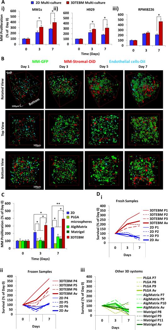
Figure 2. 3DTEBM cultures promote MM cell proliferation better than 2D and commercially available 3D systems.
A) Growth of MM cell lines i) MM1S, ii) H929, and iii) RPMI8226 in multi-culture with MM-derived stromal cells and endothelial cells in classic 2D cultures or in the 3DTEBM at days 3 and 7; (*) p< 0.02. B) Confocal microscopy images of multi-cultures of MM1s-GFP (green), MM-derived stroma-DiD (red) and endothelial cells-DiI (cyan) in the 3DTEBM at 1, 3, 5, and 7 days of culture, shown from 3 perspectives: Z-Stack rotated view, top down view, and bottom up view; Scale bar= 100 μm. C) Growth of MM1s in multi-culture with MM-derived stromal cells and endothelial cells in classic 2D cultures, PLGA microspheres, AlgiMatrix, Matrigel, and inside 3DTEBM at days 3 and 7 compared to day 0, (*) p< 0.01, (**) p< 0.05. D) Growth of i) primary fresh CD138+ plasma cells from three MM patients, ii) primary frozen CD138+ cells from three additional MM patients, in multi-culture conditions in classic 2D cultures (blue) or in the 3DTEBM (red), iii) and primary fresh in PLGA microspheres (green), AlgiMatrix (light green), and Matrigel (dark green) at days 3 and 7; bold line reflects average growth in the different conditions.

