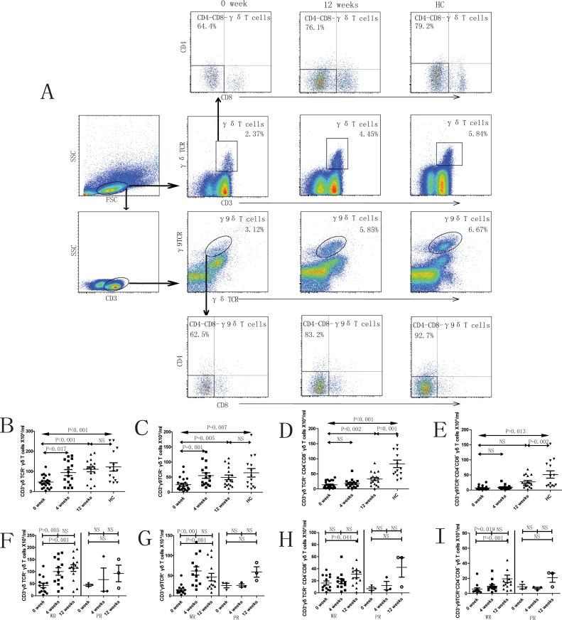
Fig 1. Peripheral blood γδ T cells and subsets in SLE patients.
A. The panels in this section show the gating strategy employed for the analysis of γδ T cells and subsets. Peripheral venous blood derived leukocytes were stained with different fluorescent antibodies and after lysis of red blood cells, the remaining cells were gated on living lymphocytes, then gated on CD3+γδTCR+ or CD3+γδTCR+γ9TCR+ cells, and then further gated on CD4-CD8- cells, respectively. B—I. Flow cytometry results are represented as the scatter dot plots of γδ T cells and subsets in healthy controls (HC) or in SLE patients before and after therapy (treatment time is indicated). SLE patients are further grouped as responders (R) and non-responders (NR) based on response to therapy.

