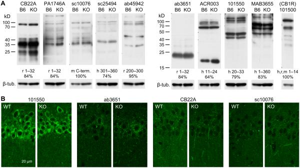
Figure 7.
Tests for the specificity of anti-CB2R antibodies. A. Western blotting tests for anti-CB2R antibodies. Nine antibodies against CB2R were tested with proteins extracted from the hippocampi of C57BL/6J (B6 lanes) and CB2R KO (KO lanes) mice. The vendors’ product numbers of antibodies are indicated above the blots. An anti-CB1R antibody (Cayman 101500) was used for the hippocampus of C57BL/6J mice as a control for GPCR extraction. Indicated below each blot are the species and amino acid sequence of the epitope for each antibody, as well as percent homology of the epitope with the corresponding mouse sequences (h, human; r, rat; m, mouse). The same membranes were used after stripping for β-tubulin detection. B. Fluorescent immunohistochemistry with anti-CB2R antibodies and hippocampal slices cut from CB2R WT and KO mice. The product numbers of antibodies are indicated above each pair of images of strata pyramidale and radiatum of the CA1 area.

