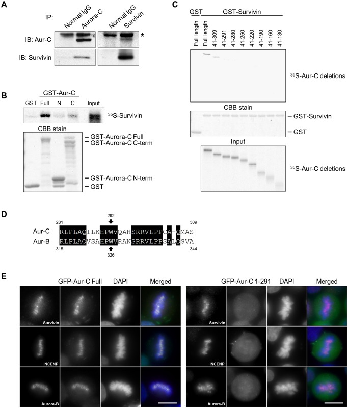
Fig 1. C-terminus end of Aurora-C binds with Survivin and is responsible for its localization.
(A) PC3 cells were treated with 500 ng/ml nocodazole for 24 h and lysed with L buffer containing 250 mM NaCl. 1 mg of lysate was immunoprecipitated with 1 μg of anti-Aurora-C, anti-Survivin, or normal IgG antibodies respectively. Immunoprecipitates were immunoblotted with indicated antibodies. The asterisk indicates non-specific bands. (B) 35S-labeled Survivin was incubated with beads bound to any of the following: GST, GST-Aurora-C full-length (Full), N-terminus amino acids 1–40 (N), or C-terminus amino acids 41–309 (C). Beads were resolved by SDS-PAGE, and visualized by autoradiography (for binding, top) or CBB stained (bottom). (C) 35S-labeled Aurora-C full length or deletion mutants were incubated with beads bound to GST or GST-Survivin, and analyzed as in B. The panel shows autoradiography (for binding, top; for input, bottom) or CBB stained gel (middle). (D) Alignment of the C-terminus sequences of Aurora-C and Aurora-B. (E) Localization of GFP-Aurora-C full length or GFP-Aurora-C deletion mutant (amino acids 1–291) in HeLa cells. Transfected cells were immunostained with indicated antibodies (red). DNA was stained with DAPI (blue). Scale bars indicate 10 μm. Each experiment was repeated at least twice with two to three biological replicates and the figures show representative images.

