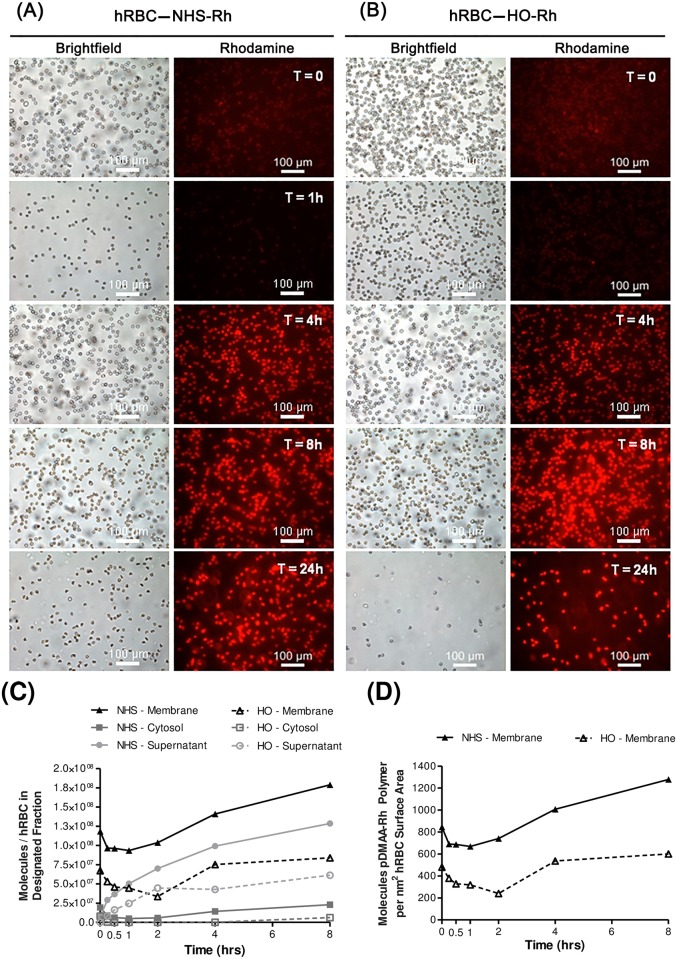
Fig 2. Human erythrocyte membrane engineering with NHS-pDMAA-Rh polymers.
NHS-pDMAA-Rh polymers exhibit significant non-specific binding and initial high-density fluorescence quenching. Human RBCs were modified with 182 μM NHS-pDMAA-Rh or 98 μM HO-pDMAA-Rh for 30 minutes at 37°C. (A) Representative images of NHS-pDMAA-Rh-exposed hRBC for each designated time point. (B) Representative images of HO-pDMAA-Rh-exposed hRBC for each designated time point. Epifluorescent images were capture after washing on a Leica inverted microscope at 20X. Scale bars measure 100 μm. (C) Supernatant, cytosolic, and membrane fractions were collected at 0, 1, 4, and 8 hours. Polymer retention and internalization was assessed by monitoring the relative fluorescence of each fraction over time and calculated as the number of polymer molecules per hRBC using a standard curve. (D) The number of polymer molecules per nm2 hRBC surface area was calculated using an average hRBC surface area of 140 μm2. Images were background corrected and the brightness/contrast for each channel was balanced using Image J software. n = 3.

