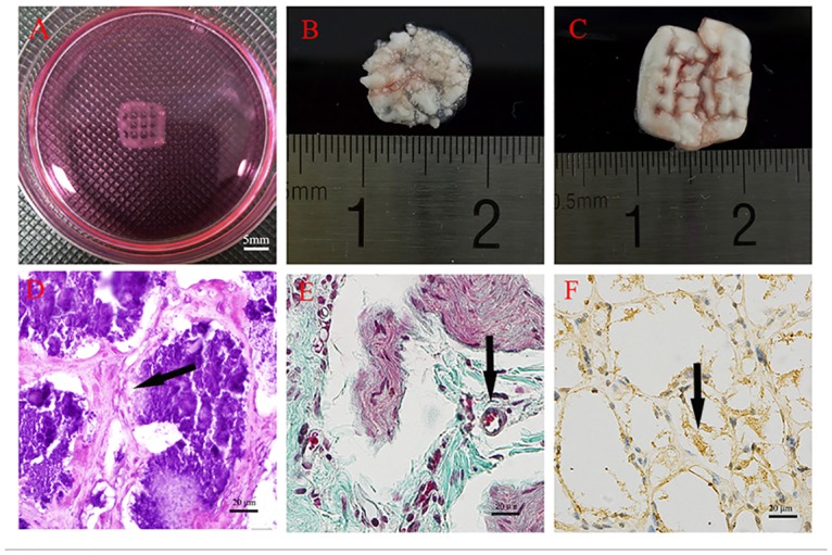
Fig 11. Ectopic bone formation.

A: The 3D bioprinted construct before implantation. B: The degraded material without hASCs. C: The 3D bioprinted construct containing hASCs. D: H&E staining of paraffin-embedded sections at 8 weeks after implantation showing obvious new bone matrix formation. E: Masson trichrome staining showing new bone matrix formation. The arrow indicatesblood vessels. F: OCN immunohistochemical staining showing obvious OCN expression in the surrounding cells (see S4 and S5 Figs).
