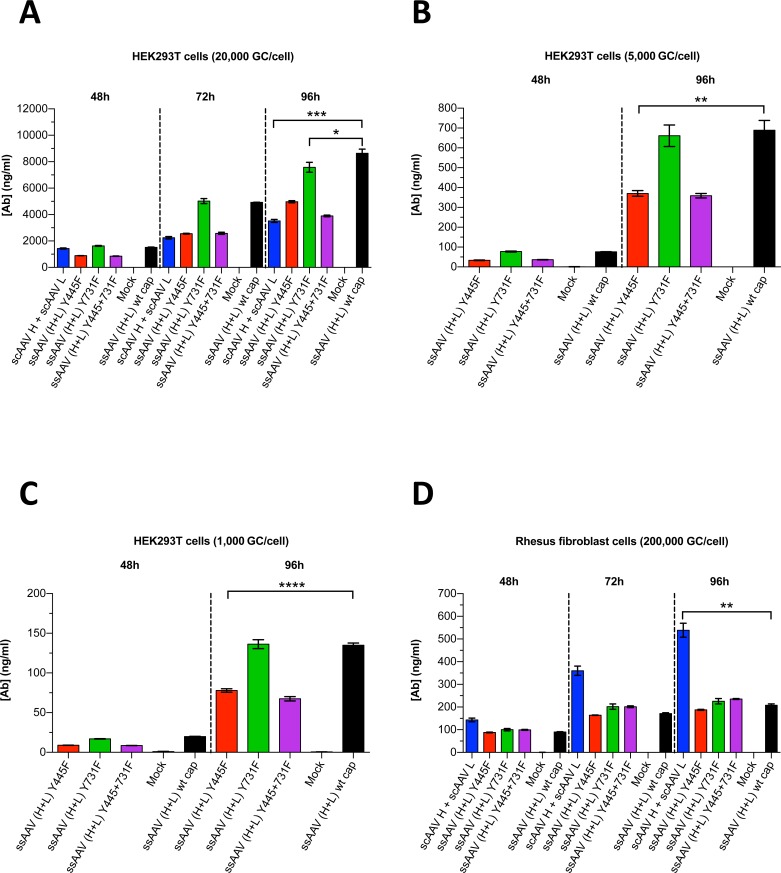
Fig 7. Expression of 5L7 IgG1 after AAV-mediated transduction in vitro.
AAV vectors were encapsidated with AAV1 wild-type (wt) capsid or AAV1 mutant capsids (Y445F and/or Y731F); in the case of ssAAV, we utilized the modified ssAAV vector construct containing both SGSG and WPRE. Purified AAV virus particles were then used for transduction. HEK293T cells were infected with (A) 2x10⁴ rAAV genome copies per cell (GC/cell), (B) 5x10³ GC/cell and (C) 1x10³ GC/cell. (D) Rhesus fibroblast cells were infected with 2x10⁵ GC/cell. AAV transduction experiments shown in (A + D) were conducted at a different time than experiments in (B + C). Levels of secreted antibody were measured by ELISA following the time of transduction. Values are depicted as mean ± SD (n = 3/group); ****p < 0.0001, ***p < 0.001, **p < 0.01, *p < 0.05.

