
Figure 1.
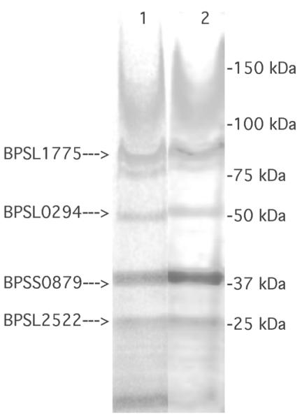
SDS-PAGE and PMF analysis of purified OM from B. pseudomallei. Ten migrograms (lane 1) and 40 μg (lane 2) of protein from a Triton-insoluble OM pellet that had been further purified by extraction with RNase, Na2CO3, and NaCl were boiled for 5 min in denaturing solution (10 mM Tris-HCl pH 6.8, 2% SDS, 2% mercaptoethanol) and electrophoresed at 100 V for 3 h on a 4 to 20% polyacrylamide gradient gel (Thermo Fisher 25204). After Coomassie Blue staining, bands were excised and the protein species present in them were identified by PMF. Locus tags of PMF-identified OMPs are on the left; migration of molecular weight markers is shown on right. Cells were grown in medium 2.