
Fig. 2.
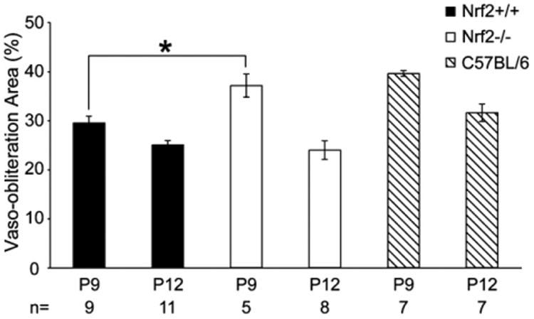
The percentage of retina with vaso-obliteration in the lectin-stained retinas after two (P9) and five days (P12) of hyperoxia. The mean percent area of retina with vaso-obliteration in each group was compared between Nrf1−/− and Nrf1+/+ mice at each time point by unpaired t-test. Asterisk indicates P < 0.05 between Nrf+/+ and Nrf−/− mice at P9 and the bars indicate SEM.