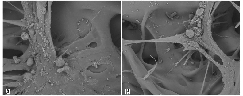
Figure 7.
Observation of chitosan porous scaffold and BMSCs co-culture for 48 h under scanning electronic microscope. (A) Wide spread of BMSCs on the surface and pores of the scaffold; (B) Bundle of cells with secreted extracellular matrix on the scaffold, with the pseudopods of some cells stretching out into the scaffold.

