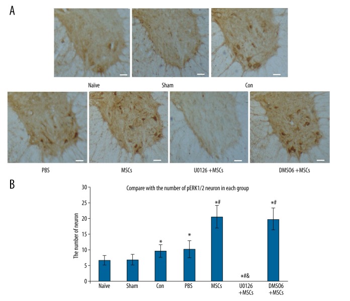
Figure 5.
Effect of intrathecal transplantation of MSCs on the numbers of pERK1/2 neurons in the spinal cord following ischemia-reperfusion injury. (A) The immunohistochemical staining of pERK1/2 neurons in the spinal cord slice in every group. Scale bar=50 μm. (B) The statistical analysis of the number of pERK1/2 positive neurons in every group. Data are shown as the mean ±SEM (n=8). * P<0.01, vs. naïve group, # P<0.01 vs. con group, & P<0.01, vs. MSCs group.

