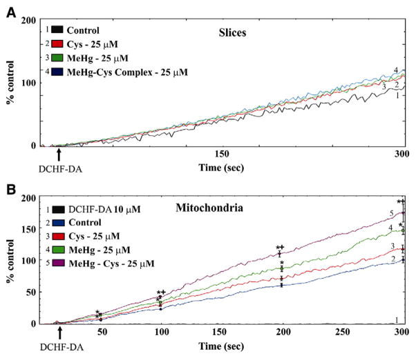
Fig. 3.
Effects of exposure to MeHg or the MeHg–Cys complex on DFC-RS production in rat liver slices (A) and mitochondria (B). Slices were exposed for 30 min to MeHg (25 μM) or the MeHg–Cys complex (25 μM each). (*Indicates p<0.05 from control; +Indicates p<0.05 from MeHg; n=5 mean±S.E.). The tracings of Figs. 3A and B are representative and averaged lines respectively.

