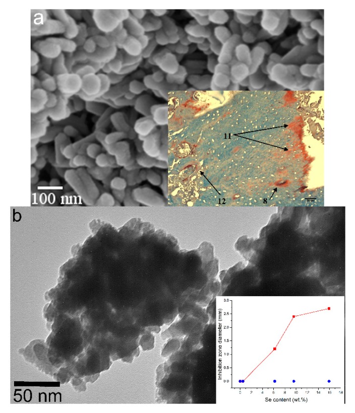
Figure 5.
(a) SEM image of hydroxyapatite nanoparticles in which Ca2+ ions were partially substituted with Co2+ ions. The inlet shows an almost complete resorption of the implant and the regeneration of an osteoporotic bone 24 weeks after the implantation (8—formation of Haversian canals; 11—ossification frontline; 12—collagen fibers; all in-between is the region filled by the newly regenerated bone); (b) TEM image of selenite-incorporating hydroxyapatite nanoparticles. The inlet shows the diameter of E. coli growth inhibition zone around particles of selenite-doped hydroxyapatite as a function of the concentration of selenite ions inside hydroxyapatite particles and depending on whether the selenite ions were incorporated into the lattice by co-precipitation () or by ion-exchange sorption ().

