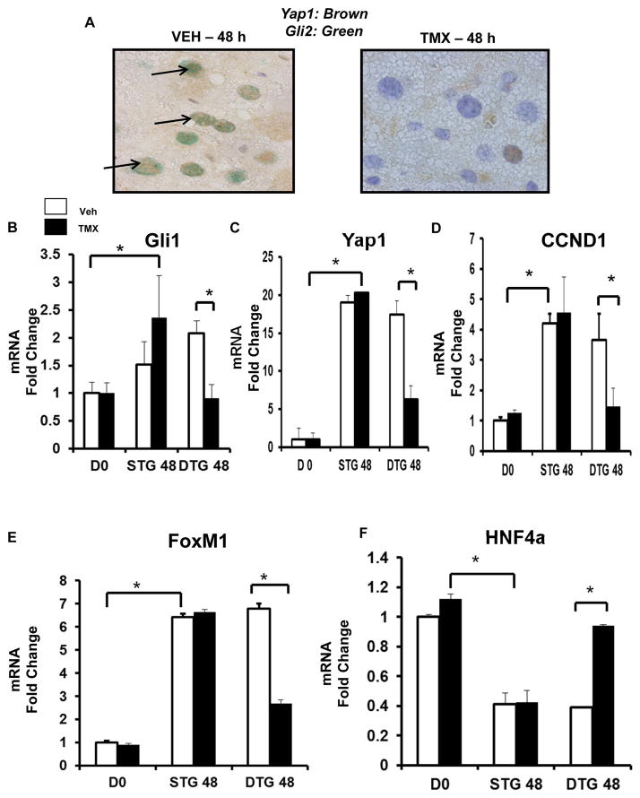
Figure 6. Hedgehog pathway activity in αSMA (+) cells promotes proliferation and de-differentiation in hepatocytes.
αSMACreERt2/Smoflox/flox (DTG) mice were treated as described in Figure legend 5. Livers were analyzed by immunohistochemistry. (A) Yap1 (brown)/Gli2 (green) double immunostaining in VEH- and TMX-treated DTG 48 h post-PH (black arrows; 100x). In separate experiments, hepatocytes were isolated from VEH or TMX-treated DTG (n =10) and STG (n = 10) 48 h post-PH, and at time 0 (i.e. DO), and analyzed by qRT-PCR. (B) Gli1 mRNA. (C) YAP1 mRNA. (D – E) Proliferation markers: CCND1 (D) and FoxM1 (E) mRNA. (F) Hepatocyte marker, HNF4a mRNA. Results were expressed as fold change relative to DO hepatocytes; mean ± SEM were graphed; *p<0.05 versus DO or VEH-treated DTG hepatocytes.

