Figure 1.

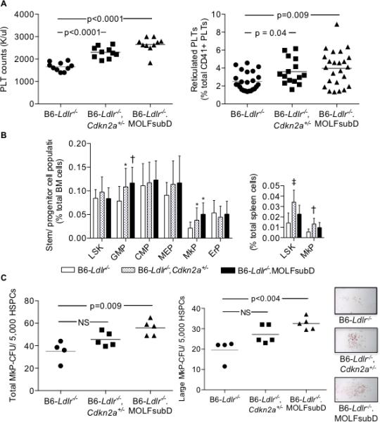
Increased PLT production in chow-fed, Cdkn2a-deficient strains in the B6-Ldlr−/− background. A: PLT counts from EDTA whole blood. Reticulated PLTs from acid-citrate-dextrose blood incubated with CD41-APC and thiazole orange nucleic acid-binding dye and analyzed by flow cytometry: B: LSK and progenitor subpopulations from BM or spleen were measured by flow cytometry. LSK (Lin−Sca1+c-kit+), GMP (Lin−Sca1−c-kit+CD34intFcγRII/IIIhi), CMP (Lin−Sca1−c-kit+CD34intFcγRII/IIIint), MEP (Lin−Sca1−c-kit+CD34intFcγRII/IIIlowCD71−CD41−), MkP (Lin−Sca1−c-kit+CD34intFcγRII/IIIintCD71−CD41+), and ErP (Lin−Sca1−c-kit+CD34intFcγRII/IIIloCD71+CD41−). N = 20 (BM) or 10 (spleen) mice/group. ‡p=0.0003, †p<0.002, and *p<0.05 vs B6-Ldlr−/− control. C: MkP colony-forming units derived from BM cells following 12 days of culture with 50 ng/ml TPO/10 ng/ml IL-3 and stained for acetylcholinesterase activity. One-way ANOVA (A, PLT counts and B); Kruskal-Wallis non-parametric (A, Reticulated PLTs and C). HSPC, hematopoietic stem and progenitor cells; HPF, high-power field.
